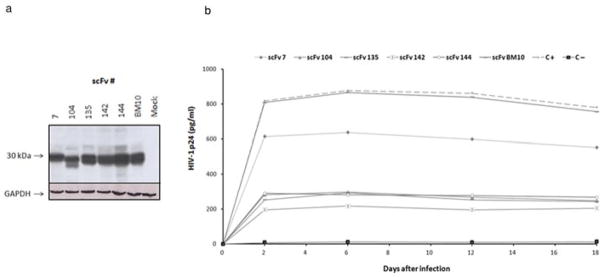
Fig. 7.

HIV-1 challenge of stable anti-IN–scFv–Jurkat cell clones. (a) Western blot of Jurkat cells stably expressing anti-IN–scFvs and anti-LANA1 scFv (clone BM10). Lysates of Jurkat cells not expressing scFv were used as negative controls (mock). Loading was controlled with anti-GAPDH antibody (Santa Cruz Biotechnology, Santa Cruz, CA, USA). (b) Stable Jurkat cell clones expressing anti-IN–scFv intrabodies were infected with HIV-1NL4-3 at a MOI of 0.1–0.5. The cultures were maintained for up to 20 days. To monitor infection, aliquots were taken at the indicated time points and p24 levels were determined by ELISA. BM10 was used as a control antibody. C + indicates HIV-1NL4-3 infected cells; C− indicates uninfected cells. The data are representative of two independent experiments.
