
Figure 4. Vav1 activation is sufficient for matrix degradation downstream of Src.
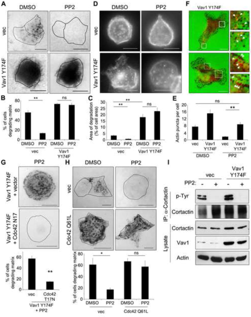
(A) DanG cells were transfected with control vector or active Vav1 Y174F, then treated with the Src inhibitor PP2 (10 μM) or DMSO vehicle control while plated on a fluorescent gelatin matrix for 7 hours. (B) The percentage of cells degrading the matrix was scored (n>100 cells per condition). (C) The area of degradation was quantified in n>20 cells per condition. Note that Vav1 Y174F potently induced matrix degradation, even in the presence of PP2. (D) Vav1 Y174F rescues the formation of invadopodia. DanG cells were transfected as described in (A), and plated on fluorescent gelatin overnight in the presence of BB94 (1 μM). The BB94 was washed out and cells were incubated for 4 hours +/- PP2 prior to staining for actin to mark invadopodia, and Vav1 to detect transfected cells (not shown). (E) The number of actin puncta per cell was scored in at least 20 cells per condition. (F) Vav1 Y174F can be detected at sites of matrix degradation. DanG cells were transfected with Vav1 Y174F, and plated on fluorescent gelatin (green) for 7 hours. Cells were fixed and stained for Vav1 (red). The boxed regions are magnified at right. Arrows indicate Vav1 Y174F puncta that colocalize with sites of matrix degradation. (G) DanG cells virally transduced to stably express Vav1 Y174F were transfected with empty vector or dominant negative Cdc42 (T17N, myc-tagged), then plated on fluorescent gelatin in the presence of 10 μM PP2. Overexpressed Vav1 and Cdc42 T17N were identified by immunofluorescence for Vav1 and myc, respectively (not shown). Cdc42 T17N inhibits matrix degradation induced by Vav1 Y174F. (H) DanG cells were transfected with myc-tagged active Cdc42 (Q61L) or empty vector, and plated on fluorescent gelatin +/- PP2 (10 μM) for 7 hours. Cdc42 Q61L expression was verified by anti-myc immunofluorescence (not shown). Cdc42 Q61L was sufficient to rescue matrix degradation in the presence of PP2. For E and F, the percentage of cells degrading matrix was scored in at least 50 cells per experiment. (I) The Src substrate cortactin is not phosphorylated upon PP2 treatment. DanG cells stably expressing GFP vector or Vav1 Y174F were treated with PP2 (10 μM) or DMSO control, then immunoprecipitated for cortactin and blotted for phospho-tyrosine. Note that cortactin is not phosphorylated in the PP2-treated cells, even in the cells expressing Vav1 Y174F. All graphed data represent the mean +/- SEM of three independent experiments. * indicates p<0.05, ** indicates p<0.01, and ns indicates no statistically significant difference. Scale bar = 10 μm. See also Supplemental Figure S4.