
Figure 2.
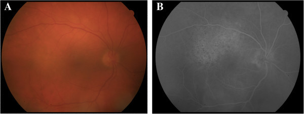
Patient 2, pretreatment fundus imaging of the affected eye. (A) Fundus photograph demonstrating broad areas of deep retinal whitening along the superotemporal and inferotemporal arcades. (B) Early phase fluorescein angiogram with leopard spotting in the superior macula.