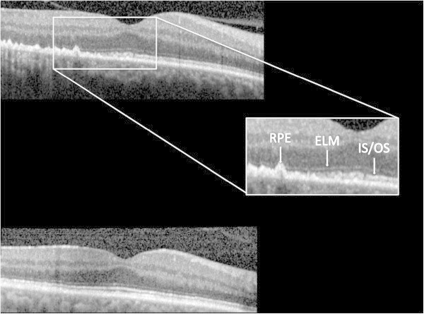
Figure 5.

Spectral domain optical coherence tomography images from the affected eye of patient 2. The top image shows pre-treatment pathology, including (inset) retinal pigment epithelium (RPE) nodularity, disruption of the external limiting membrane (ELM), and loss of the photoreceptor inner segment/outer segment (IS/OS) band. The bottom images demonstrate resolution of these changes after antibiotic therapy.
