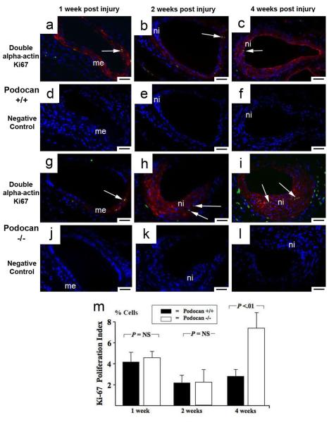
Figure 3.
Arterial proliferation with wild type and podocan−/− genotype. Ki-67 (FITC) and smooth muscle alpha-actin (Texas Red) double labeling: WT genotype (a-f): podocan−/−genotype (g-l): 1 week (a and g): Early after injury Ki-67 positive (green) and alpha-actin positive (red) SMC (arrow) are seen in the media (me) in both groups; x200; scale bar=50 μm. (d and j represent matching IgG-isotype control stainings). 2 weeks (b and h): At this time only few Ki-67 signals (arrows) are seen in both groups consistent with a gradual decline in proliferation after the first week; x200; scale bar=50 μm. (e and k negative controls). 4 weeks (c and i): An unusually late rise in proliferation of SMC (red alpha-actin labeling) is detected by nuclear Ki-67 (green) labeling (arrows) with podocan−/− genotype; x200; scale bar=50 μm. (f and l negative controls). Bar graph (m): Comparison of Ki-67 expression with WT and podocan−/− genotype after arterial injury: expression in % cells (independent sample t-test).

