Figure 5.
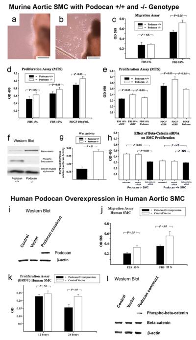
Effect of WT and podocan−/− genotype on murine aortic SMC: Aortic explant culture (a and b): (a) The edge of WT aortic explant shows no SMC outgrowth at day 3; x400; scale bar=50 μm. (b) In contrast, numerous SMCs are seen at the edge of a podocan−/− aortic explant at the same time point; x400; scale bar=50 μm. SMC migration and proliferation (c-e): (c) SMC migration is increased with podocan −/− genotype compared with WT in a colorimetric test based on the Boyden chamber principle (independent sample t-test). (d) Podocan−/− SMCs also grow faster as measured by the MTS assay (independent sample t-test). (e) Podocan−/− SMC transfected with podocan vector slow their growth to WT-level (independent sample t-test). Wnt-TCF pathway activation (f-h): (f) In SMC with podocan−/− genotype the ratio of phosphorylated to non-phosphorylated beta-catenin is reversed as seen by Western blot. (g) Transcriptional activity of Wnt-TCF pathway measured directly by TOPflash/FOPflash assay is also increased with podocan−/− genotype. (h) Podocan−/− SMCs treated with small inhibitory RNAs to beta-catenin show inhibition of growth. Effect of podocan overexpression on human aortic SMC: Western Blot (i): Western Blot confirms overexpression of the human form of podocan. Podocan in control and empty vector treated SMC is below the detection threshold. Migration (j): SMC migration is reduced by 29% with podocan overexpression. Proliferation (k): SMC proliferation is reduced by 32% with podocan overexpression. Wnt-TCF pathway activation (l): SMC overexpressing podocan show an increase in phosphorylated-beta catenin on Western Blot compared to non-treated or empty vector treated SMC indicating Wnt-TCF pathway suppression.

