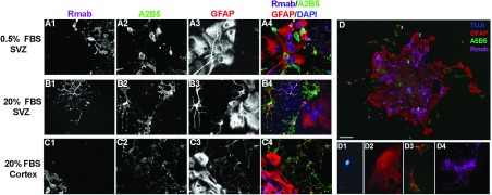
Figure 5. Spheroids from the neonatal SVZ and neocortex generate colonies that contain neurons, type 1 astrocytes, type 2 astrocytes and oligodendrocytes.

Spheroids were cultured with 10 ng/ml PDGF-AA, differentiated for 3 days in either 0.5% FBS or 20% FBS then stained for oligodendrocytes (Rmab) (A1, B1, C1), O-2A lineage (A2B5) (A2, B2, C2), and astrocytes (GFAP) (A3, B3, C3). The pseudocolor images show an overlay of Rmab (magenta), A2B5 (green), GFAP (red), and DAPI (blue) (A4, B4, C4). When differentiated in 0.5% FBS and 10 ng/ml BMP-4, a single spheroid produced neurons (TUJ1), type 1 astrocytes (A2B5−/GFAP+), type 2 astrocytes (A2B5+/GFAP+) and oligodendrocytes (Rmab) (D). Examples of the four cell types from a single spheroid neuron (D1), type 1 astrocyte (D2), type 2 astrocyte (D3) and oligodendrocyte (D4) within the spheroid. Scale bar represents 20 μm.
