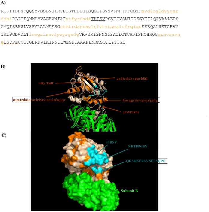
Figure 4. The predicted epitopes of Stx2 to antibody 11E10.
A) The predicted epitopes of Stx2 to antibody 11E10 are highlighted in lower case and colored orange in the protein sequence. The recognition regions identified previously (Smith et al 2009) are underlined. B) Backbone presentation of the antigen subunits A and B showing the predicted epitopes in orange and identified regions A-C colored in blue. The antibody binds to subunit A only. Subunit B is shown in green. C) Surface presentation of the antigen subunits A and B showing the predicted epitopes in orange and recognition regions A, B and C colored in blue. Note that the region C is only partially shown as the region is missing in the crystal structure of Stx2 (PDB 1R4P) (Fraser et al, 2004). Figures were prepared using PyMOL [38].

