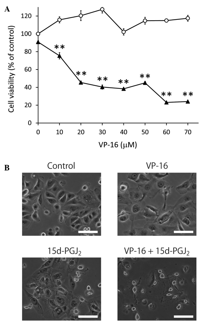
Figure 2.

Treatment with VP-16 enhanced the antiproliferative effect of 15d-PGJ2 in Caki-2 cells. (A) The cells were assayed for viability using MTT following treatment with VP-16 (0, 10, 20, 30, 40, 50, 60 and 70 μM) (open circles) and combination treatment with VP-16 and 15d-PGJ2 (20 μM) (closed triangles) for 24 h. The results are expressed as the means ± SEM of three independent experiments. **P<0.01, vs. control cells. (B) The combination of 15d-PGJ2 and VP-16 induced morphological changes in Caki-2 cells. The cells were treated with 15d-PGJ2 alone (20 μM), VP-16 alone (70 μM) and the combination of the two. Caki-2 cells were then examined by phase contrast microscopy following 24 h of incubation. Scale bar, 100 μm. 15d-PGJ2, 15-deoxy-Δ12,14-prostaglandin J2; VP-16, etoposide.
