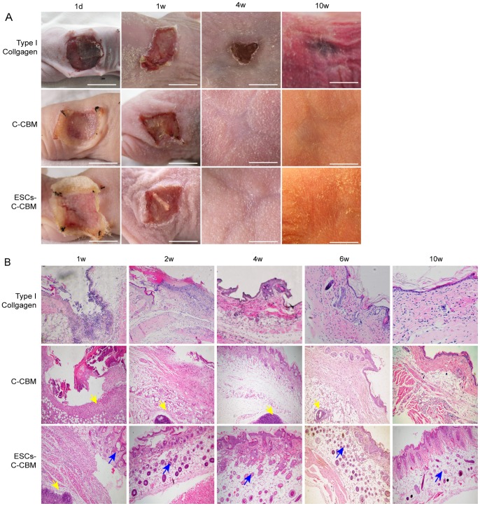
Figure 3. Observation of nude mice full-thickness defect model.
(A) Observation of nude mice full-thickness defect model dressed with type I collagen only, C-CBM, or ESCs-C-CBM at 1 d, 1 w, 4 w, and 10 w. Type I collagen group shows that wounds are much slower to heal. Group C-CMB shows the repaired wound skin in the control group was relatively thin and heliotrope with a tendency to bleed; Group ESCs-C-CMB shows the repaired wound in the experimental group was relatively thick and red with re-epithelialization. Scale bar = 1cm. (B) Formation of epidermal nests on the wound surface repaired by epidermal stem cells- collagen-chitin biomimetic (ESCs-C-CBM) membrane compared with C-CBM. Paraffin and stained with hematoxylin and eosin (H&E) stain. Yellow arrows point to chitin and blue arrows point to epidermal nests increased in the ESC-C-CBM group at 2–10 w (100X).

