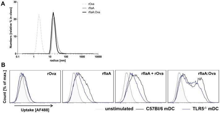
Figure 3. rflaA:Ova forms aggregates which are efficiently internalized by mDC.
Hydrodynamic radii of rOva, rflaA and rflaA:Ova were determined via lightscattering (A). Uptake of Alexa Flour 488 labelled proteins into C57Bl/6 wt (black) and TLR5−/− (blue) CD11c+CD11b+B220- mDC stimulated with rOva (1 µg/ml), rflaA (0.7 µg/ml), rflaA (0.7 µg/ml)+rOva (1 µg/ml) or rflaA:Ova (1.7 µg/ml) was quantified 15 min post stimulation via flow cytometry (B).

