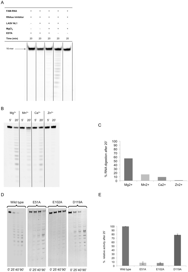
Figure 3. In vitro endonuclease activity of LASV L173.
A, A 5′ FAM-labeled 16-nt single-stranded RNA substrate was incubated in a buffer with or without purified LASV endonuclease, with or without Mg2+ and with or without metal ion chelator EDTA, for 20 min at 37°C. The reaction products were separated by urea-PAGE and detected by fluorescence scanning. B, The in vitro endonuclease assay was conducted in buffers with different divalent cations for either 5 or 20 min. C, Percentage of RNA substrate degradation after 20 min incubation in a buffer with different divalent cations was quantified by fluorescence scanning. D, WT or mutant LASV L173 was analyzed by an in vitro endonuclease assay for 0, 25, 40 and 90 min. E, Percentage of RNA substrate degradation after 25 min was quantified by fluorescence scanning and normalized to WT control (set at 100%).

