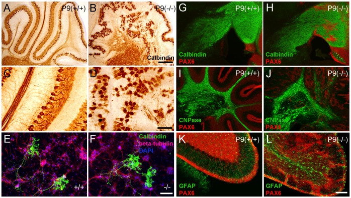
Figure 2. Cxcr4 deficiency inhibits Purkinje cell dendritogenesis caused by ectopic granule cells migration in the developing brain.
(A,B) Calbindin staining revealed ectopic and disorganized Purkinje cells in P9 KO mice. (C,D) A greater magnification of figure A and B shows Purkinje cells from P9 KO mice have less dendritogenesis and feature axon disorganisation (n = 6). (E,F) Cerebellum cell culture from E17 KO and WT embryos. Cabindin staining (green) shows there is no significant difference in the dendritic development of Purkinje cells between KO and WT cells (n = 12). No obvious defect in Purkinje cell axon growth was observed. (G,H) Calbindin staining reveals the axons of Purkinje cells project to the deep cerebellar nuclei correctly in KO mice. (I,J) In P9 WT mice, we observed compact myelinated axon fibers by staining CNPase. In KO mice these myelinated fibers are disorganized and sporadic. (K,L) Pax6-expressing granule cells migrate along the GFAP-positive radial glia fibers in WT mice. In KO mice, the GFAP positive cells do not align neatly in the Molecular Layer and the Pax6-expressing granule cells do not migrate appropriately. Scale bars = 300 µm in A,B; 100 µm in E,F. 200 µm in G,H,I,J. 100 µm in K,L.

