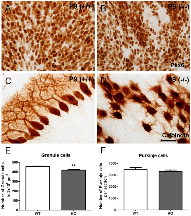
Figure 3. Cxcr4 deficiency results in lower density of granule cells, but no difference in the number of Purkinje cells in P9 cerebella.
(A,B) The Pax6 staining shows the granule cells in the WT and KO P9 cerebella. (C,D) The Calbindin staining shows the disorganized Purkinje cells in KO cerebellum. (E,F) There is lower density of granule cells in KO cerebella, but no difference in the number of Purkinje cells. Values represent mean ± SEM (** p<0.01). Scale bar = 30 µm in A,B,C,D.

