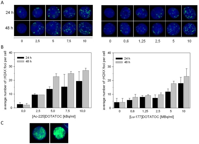
Figure 3. Number of γH2AX foci in AR42J cells at 24 and 48 h after incubation with 225Ac-DOTATOC (left) and 177Lu-DOTATOC (right).
(A) shows representative images from all activity levels, (B) shows quantification of γH2AX foci and (C) shows two representative examples for pan nuclear staining after high dose 225Ac-DOTATOC treatment.

