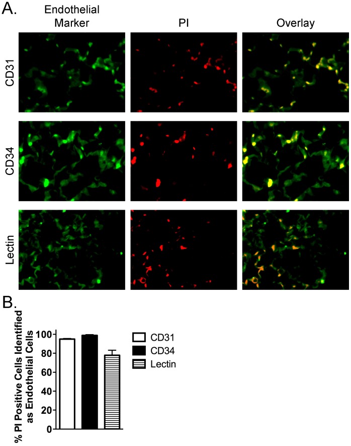
Figure 5. Confirmation that CLP/sepsis-induced non-viable (PI-positive) pulmonary cells are pulmonary microvascular endothelial cells.
(A) Endothelial cells (left column of panels; green) were labeled with specific markers, including CD31 (PECAM; top left panel), CD34 (middle left panel), and lectin (lower left panel) in histologic pulmonary sections. Nuclear PI (red) staining identifies non-viable cells (middle column of panels). Overlap between cells positive for endothelial markers and PI (yellow) specifically identifies non-viable endothelial cells (right column of panels). (B) Quantification of the percentage of PI-positive cells that also stain positive for markers of endothelial cells confirms that the majority of CLP/sepsis-induced PI-positive/non-viable pulmonary cells are MVEC. All images 63X.

