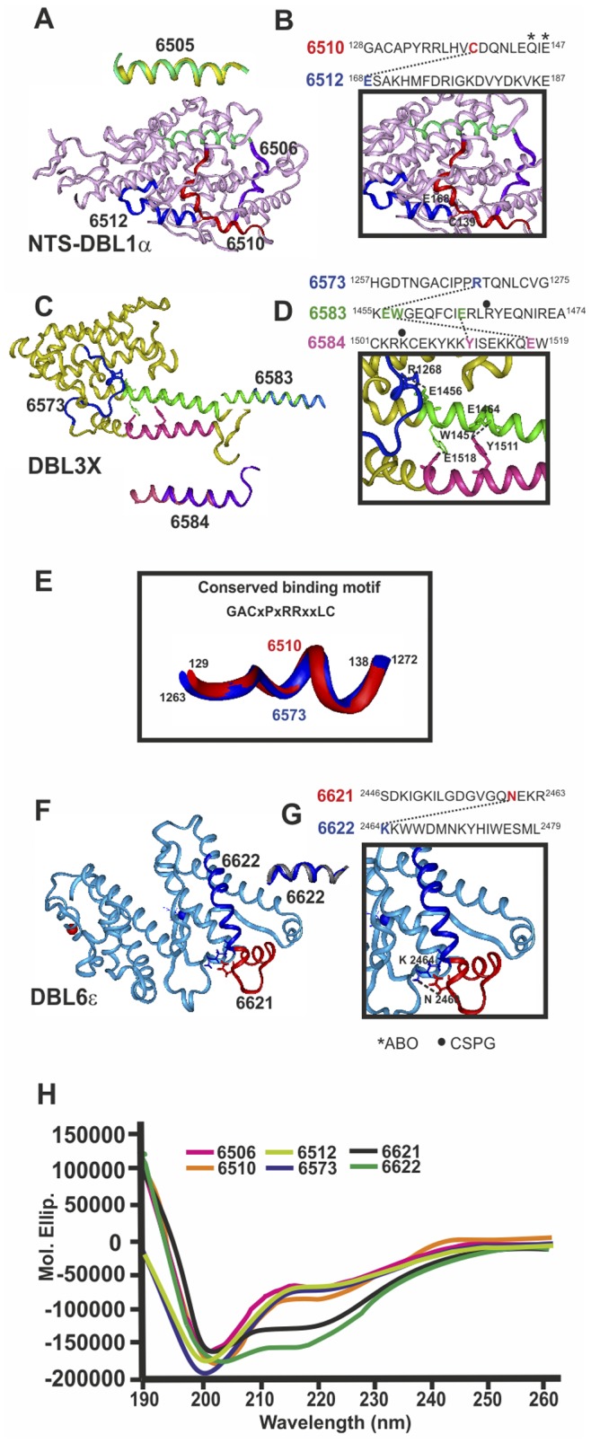
Figure 4. Structural characterization of HABPs present in crystallized Duffy binding like domains (DBL).

DBL domain 3D structure determined by X-ray crystallography A) Head structure: DBL1α (PDB 2XU0) (pink), C) DBL3X (PDB 3CML) (yellow), F) DBL6ε (PDB 2WAU) (pale blue). 1H-NMR-determined structure localisation, displaying the perfect fit of HABP 6505 (yellow) superimposed onto DBL1α, 6583 (dark blue) and 6584 (purple) onto DBL3X and 6622 (grey) onto DBL6ε. B, D, G). H-bonds between HABP residues and their corresponding sequence on top, displaying relevant residues in binding to A blood group trisaccharides and CSPG (asterisk and black dot, respectively). E) Superimposed conserved binding motif fragments from 6510 and 6573. H) CD spectra for corresponding HABPs.
