Figure 1.

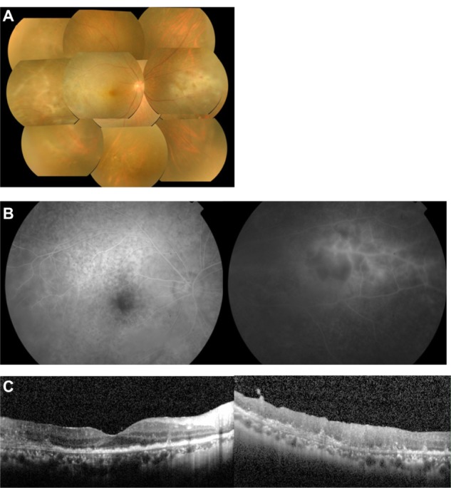
Case 1. Fundus photographs, fluorescein angiograms, and spectral-domain optical coherence tomographic images of the right eye in a 57-year-old man with primary intraocular lymphoma after vitrectomy.
Notes: (A) Color fundus photograph shows diffuse retinal infiltration in the macula and retinal vasculitis in the temporal and nasal areas. (B) Late phase fluorescein angiogram showing hypofluorescent spots with a leopard spot pattern in the posterior fundus and staining of the arteries and an avascular area in the nasal region. (C) Spectral-domain optical coherence tomographic images. (Left) Nodular hyper-reflective infiltration at the level of the retinal pigment epithelium, separation of Bruch’s membrane from the retinal pigment epithelium, partial damage of the retinal pigment epithelium, disruption of the photoreceptor inner segment/outer segment junction, and hyper-reflective signals in the inner retina can be seen. (Right) A layered structure of the retina cannot be detected on the temporal side.
