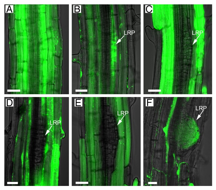
Figure 1. Symplastic connectivity between the outer tissues and the primordia is regulated during lateral root development. A symplastic fluorescent dye (green; FDA), generated from CFDA applied exogenously, moved freely through cell layers in non-lateral root regions and into stage I–II primordia (A-B). Movement was blocked after transition from stage III to IV primordia (C-E) and restored just before emergence (F). Images in the brightfield channel (superimposed) were used to distinguish the lateral root primordia (LRP, arrowed in B-F). Scale bars: 20 µm.

An official website of the United States government
Here's how you know
Official websites use .gov
A
.gov website belongs to an official
government organization in the United States.
Secure .gov websites use HTTPS
A lock (
) or https:// means you've safely
connected to the .gov website. Share sensitive
information only on official, secure websites.
