
Figure 3.
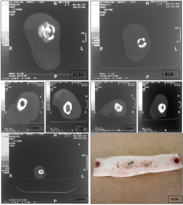
Tomography analyses during bone healing, after stem cell therapy, performed in the medial plane of the grafts of animals euthanized at 0, 6, 10, 14 and 18 weeks after the correction of diaphyseal segmental defects with cellularized homologous (A) and noncellularized homologous (B) grafts. The holes corresponding to the holes in the compact cortical bone are apparent in both animals. (Figure 3A to 3G). Gross piece of bone corresponding to the macroscopic analyses of the AOC 18 + animal (3H).