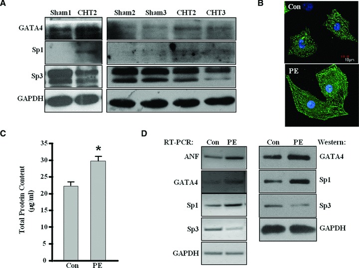
Fig 1.

The expression of GATA4, Sp1 and Sp3 in rat cardiac hypertrophic tissues and PE-treated cardiomyocytes. (A) Cardiac tissues were collected from eight hypertrophy rats and four sham rats individually. Western blot analysis was performed on individual tissues to analyse the changes of GATA4, Sp1 and Sp3 at protein level in cardiac hypertrophic rat tissue compared with control rats. The protein level of GAPDH was used as an internal control. The figure shows a representative result with three CHT rats as well three control rats. (B) Neonatal rat cardiomyocytes were cultured with phenylephrine (PE, 10 μM for 24 h) and then stained with α-actinin antibody for immunofluorescence. The cardiomyocytes cultured without PE were used as a control (Con). The experiment was repeated at least three times, the figure shows a representative result. (C) The total protein content of cardiomyocytes treated as in (B) was determined using BCA protein assay kit. (Each bar represents mean ± S.D. from three independent experiments; *P < 0.05 versus control.) (D) RT-PCR and Western blot were performed with cardiomyocytes treated as in (B) to analyse the expression of Sp1, Sp3, GATA4 and ANF. GAPDH was used as internal controls. The experiment was repeated at least three times and similar results were obtained each time. The figure shows a representative result.
