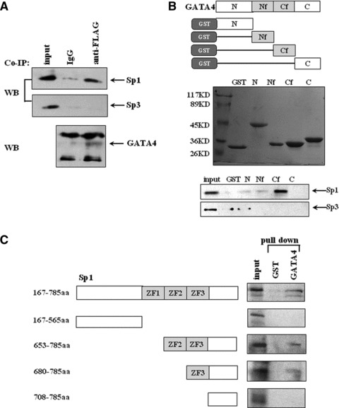
Fig 3.

Physical interaction between Sp1/Sp3 and GATA4. (A) Co-immunoprecipitation was performed with cell lysates from HeLa cells transfected with FLAG-GATA4 in combination with Sp1 or Sp3 with anti-FLAG antibody or normal IgG as a control antibody, and then detected by anti-Sp1, Sp3 or GATA4 antibodies. (B) GST pull-down experiments were performed with cell lysates from HeLa cells transfected with Sp1 or Sp3 in combination with various fragments (as shown in the upper panel) of GATA4 fused to GST and then analysed by SDS-PAGE and Western blot. N, Nf, Cf and C stand for N terminal, N terminal zinc finger, C terminal zinc finger and C terminal domain, respectively. The middle panel shows GST-fusion proteins stained by Coomassie brilliant blue. The precipitated complexes were detected with Sp1 or Sp3 antibodies (the bottom panel). An equal amount of GST protein was used as a negative control. (C) GST pull-down was performed to analyse the interaction between in vitro translated 35S-labelled various fragments of Sp1 and GATA4 fused to GST. The labelled fragment bond with GATA4 was visualized by autoradiography. All the experiments were repeated at least three times.
