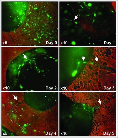
Fig 5.

Representative images showing the dynamics of GFP-labelled EPCs integration and vascular tube-like structures formation in living murine embryonic ventricular slice preparations – day 1 to day 5 of coculture. Carl Zeiss Axiovert 10 microscope and Sony Electronics Digital Interface DFW-x700 camera system.
