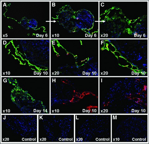
Fig 6.

Immunohistochemical analyses showing EPCs integration and vascular tube-like structures formation in living murine embryonic ventricular slice preparations. Human vWF and human nuclei expression – day 6 (A–C), day 10 (D–F), and day 14 (G) of coculture; polyclonal rabbit anti-human vWF-Alexa Fluor 488 (green), monoclonal IgG1 mouse anti-human nuclei-Alexa Fluor 555 (red), and Hoechst (nuclei, blue); negative controls-secondary antibodies only (J) and murine ventricular slice only (K). Human CD31 expression-day 10 of coculture (H, I); monoclonal IgG1 mouse anti-human CD31-Alexa Fluor 555 (red) and Hoechst (nuclei, blue); negative controls-secondary antibodies only (L) and murine ventricular slice only (M). Carl Zeiss Axiovert 200M ApoTome and AxioVision 4.6 software.
