Fig 1.

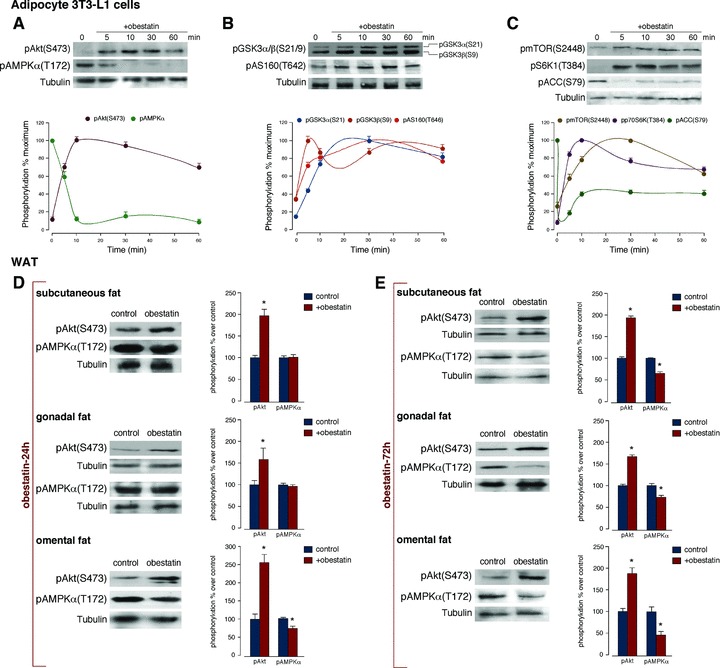
Obestatin activates Akt phosphorylation and AMPK dephosphorylation in 3T3-L1 adipocyte cells and WAT. Time-course of the effect of obestatin (100 nM) on: (A) pAkt(S473) and pAMPKα(T172); (B) pGSK3α/β(S21/9) and pAS160(T642) and (C) pmTOR(S2448), pS6K1(T384) and pACC(S79). Phosphorylation was expressed as a percentage of the maximal phosphorylation obtained for each residue (n = 3; mean±S.E.). Blots are representative of three independent experiments. (D) Effect of 24 hrs continuous sc infusion of obestatin (300 nmol/kg body weight/24 hrs; n = 10) on pAkt(S473) and pAMPKα(T172) from subcutaneous, gonadal and omental WAT obtained from male rats. (E) Effect of 72 hrs continuous sc infusion of obestatin (300 nmol/kg body weight/24 hrs; n = 10) on pAkt(S473) and pAMPKα(T172) from subcutaneous, gonadal and omental WAT obtained from male rats. Phosphorylation was expressed as a percentage of control obtained from 24 hrs or 72 hrs-minipump sc implanted saline group (n = 10; mean±S.E.). Asterisk (*) denotes P < 0.05 when comparing obestatin-treated group with untreated control group (saline).
