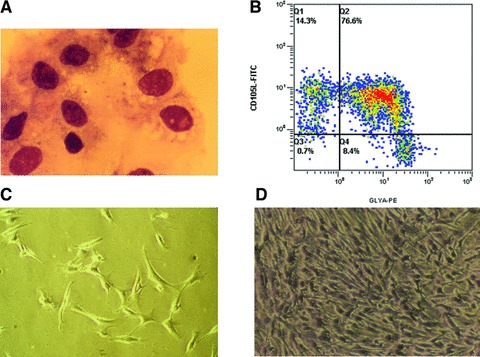
Fig 1.

Characterization of freshly isolated BM-CD105+cells and cultured MSCs. (A) Morphology of immunomagnetically isolated BM-CD105+ cells after they were cytocentrifuged and counterstained with May-Grünwald solution. (B) Three colour flow cytometric analysis of sorted cells demonstrated two distinct CD105+ cell populations: CD105+Glycophorin-A+CD45− and CD105+Glycophorin-A−CD45−. (C–D) Fibroblastoid morphology of cells derived from CD105+ cells after 3 (C) and 10 days (D) in culture before first passage (primary cells).
