
3.
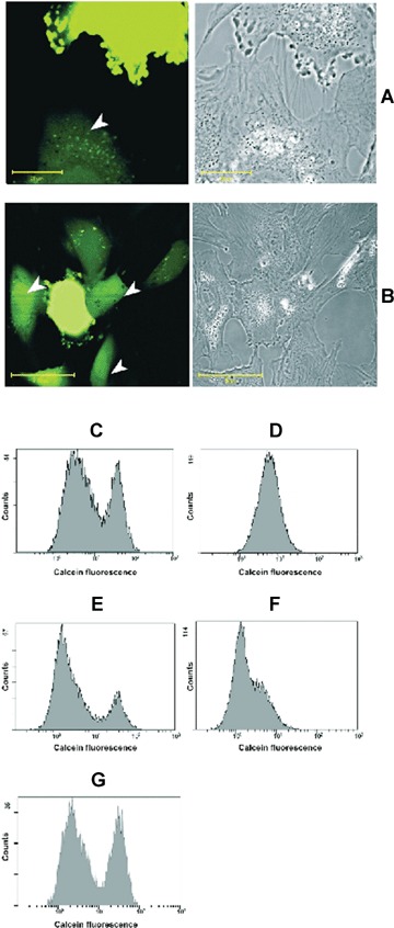
Transport of cytosolic entity from human MSC to rat cardiomyocytes after 24 hrs of co-cultivation. (A–B) MSC are stained with calcein-AM and placed to cardiomyocytes. After 24 hrs, calcein fluorescence is revealed not only in MSC but also in contacting with them cardiomyocytes (pointed with arrowheads). Diffused staining of calcein in cytosol of cardiomyocytes as well as calcein compartmentation within are observed. Calcein transfer may occur as possibly mediated by nanotubes (A) or gap junctions (B) Bar, 20 and 50 μm for A and B correspondingly. (C–G) Flow cytometry of co-cultivated human MSC and rat cardiomyocytes. Cells were stained with calcein-AM and cultivated in two varieties: C and D, MSC stained with calcein-AM while cardiomyocytes are non-stained; E and F, cardiomyocytes are stained with calcein-AM, while MSC are non-stained. Pictures are taken after 3 hrs (C and E) and 24 hrs (D and F) of co-cultivation. (G) Cells were shaken for 24 hrs on horizontal plate with 50 cycles per minute.