8.

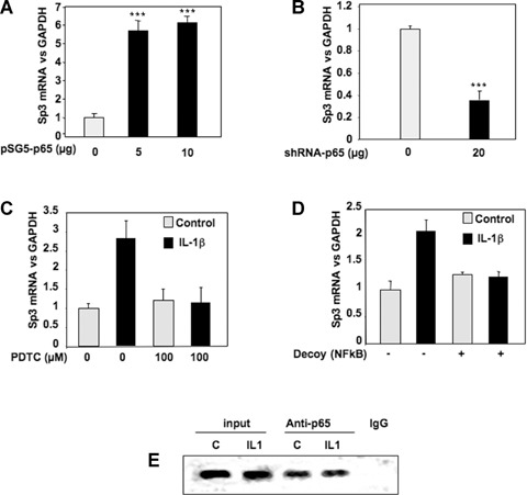
NFκB induces the expression of the repressor Sp3. A, B: Subconfluent chondrocytes were transfected with increased amounts of pSG5-p65 (A) or with 20 μg of shRNA-p65 vector (B). Thereafter, media were replaced with DMEM + 2% FCS for 24 hrs, and Sp3 mRNA levels were analysed and expressed as relative expressions versus GAPDH. C: Subconfluent cultures of chondrocytes were treated with IL-1β (1 ng/ml) for 48 hrs in the presence or in the absence of the NFκB inhibitor, PDTC. Histograms represent the relative Sp3 mRNA levels versus GAPDH signal. D: HAC were transfected with an NFκB consensus sequence (NFκB decoy). Then, they were incubated in DMEM + 2% FCS in the presence or not of IL-1R (1 ng/ml) for 24 hrs, and real-time RT-PCR analysis of Sp3 gene expression was performed. E: HAC were fixed with formaldehyde, lysed and then digested with enzyme restriction. In vivo cross-linked chromatin was then precipitated independently using anti-p65 antibody or IgG. The recovered immunoprecipitated DNA was then used for PCR with specific primers for the Sp3 promoter. The amplicons were analysed by elec-trophoresis on a 3% agarose gel.
