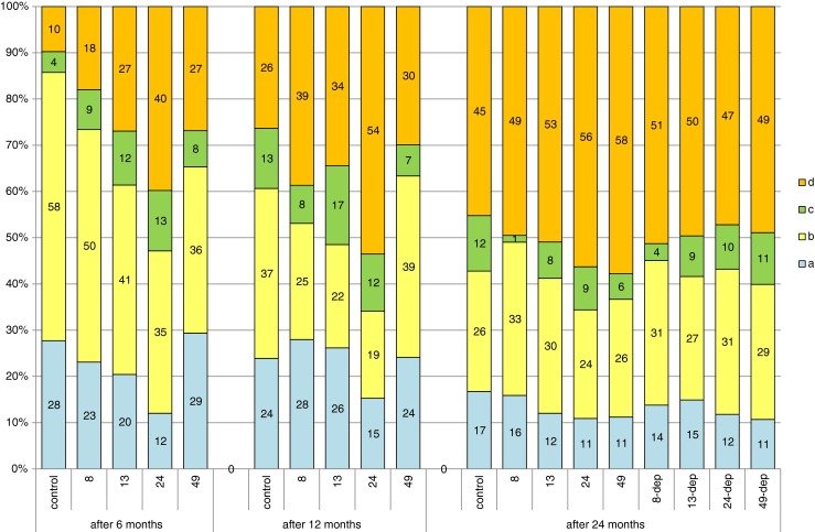
Fig. 2.

Comparison of percentages of oogonia and oocytes at different developmental stages between the groups of females (controls, 8, 13, 24, and 48 mg Pb kg−1 after 6 and 12 months of exposure and in females after 12 months of exposure and 12 months of depuration, groups 8-dep, 13-dep, 24-dep, and 48-dep) subjected to different lead exposure doses in successive years of the study: A oogonia, B oocytes in protoplasmic growth, C oocytes entering trophoplasmic growth, D oocytes in full trophoplasmic growth
