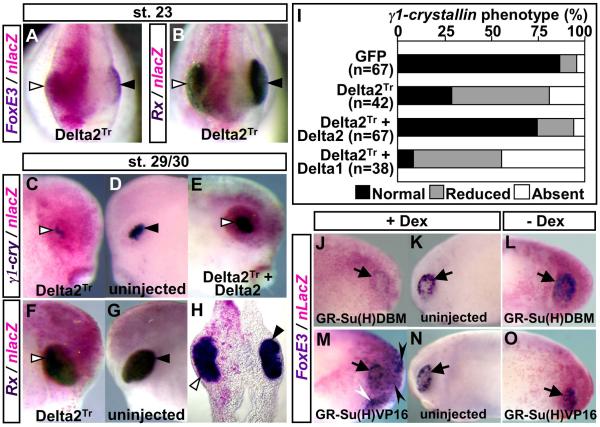
Fig. 5.
Effects of manipulation of Notch signaling on FoxE3 expression and subsequent lens placode formation. (A, B) Frontal view of embryos injected with mRNA encoding Delta2Tr (500 pg), fixed at stage 23, and subjected to lacZ staining (magenta) and in situ hybridization with FoxE3 or Rx probe (purple or deep purple staining). White and black triangles in (A–H) indicate in situ hybridization signals on injected and uninjected sides of embryos, respectively. (C–D, F–G) The injected and uninjected sides of embryos injected with Delta2Tr mRNA, fixed at stages 29/30, and hybridized with γ1-crystallin or Rx probe. (H) A transverse head section of the embryo shown in (F, G). (E) The injected side of an embryo injected with both Delta2Tr mRNA (500 pg) and wild-type Delta2 mRNA (500 pg), fixed at stage 29, and hybridized with γ1-crystallin probe. (I) Summary of Delta2Tr mRNA injection experiments. GFP mRNA (1000 pg) was injected as a control. (J, K) The injected and uninjected sides, respectively, of an embryo injected with GR-Su(H)DBM mRNA (1000 pg) and induced with Dex. Arrows in (J–O) indicate endogenous FoxE3 expression in the PLE. (L) The injected side of an embryo injected with GR-Su(H)DBM but not induced with Dex. (M, N) The injected and uninjected sides, respectively, of an embryo injected with GR-Su(H)VP16 mRNA (1000 pg) and induced with Dex. Black and white arrowheads in (M) indicate ectopic FoxE3 expression in the ectoderm overlying the anterior brain and that surrounding the cement gland, respectively. (O) The injected side of an embryo injected with GR-Su(H)VP16 but not induced with Dex.

