
Figure 4.
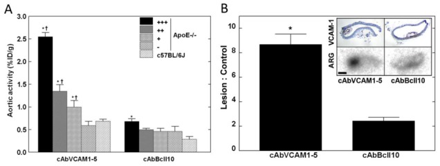
99mTc-cAbVCAM1-5 aortic distribution and autoradiography. A: 99mTc-cAbVCAM1-5 and 99mTc-cAbBcII10 aortic uptake in arterial segments from ApoE−/− mice ranked according to the lesion-extension index, and in C57Bl/6J control mice aorta. * P<0.05 vs C57Bl/6J, † P<0.05 vs next lesion-extension index. B: Representative 99mTc-cAbVCAM1-5 and 99mTc-cAbBcII10 autoradiograms (ARG) are presented together with VCAM1 immunostainings obtained on adjacent slices, showing hot-spot uptake of 99mTc-cAbVCAM1-5 in VCAM1-positive lesions. Scale bar: 200μm. (* P<0.05 vs. cAbBcII10).