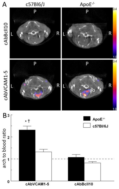
Figure 5.

99mTc-cAbVCAM1-5 as tracer for SPECT/CT in vivo imaging of atherosclerotic plaques. A: Representative in vivo SPECT/CT coronal views taken at the level of the aortic arch of C57Bl/6J and ApoE−/− mice 2–3h after i.v. injection of 99mTc-cAbBcII10 or 99mTc-cAbVCAM1-5 nanobodies. The scale was adjusted from 1 to 3.4 percent of the injected dose to allow direct visual comparison. Focal uptake of 99mTc-cAbVCAM1-5 was visible in the axillary lymph nodes (ln) and thymus (t) of both C57Bl/6J and ApoE−/− mice. In addition, 99mTc-cAbVCAM1-5 uptake in atherosclerotic lesions from ApoE−/− mice was also clearly identifiable at the level of the aortic arch (ao). B: In vivo determination of arch-to-blood ratios based on SPECT image quantifications. This ratio was significantly higher in atherosclerotic ApoE−/− than control C57Bl/6J mice for 99mTc-cAbVCAM1-5 but not for the negative control 99mTc-cAbBcII10. (* P<0.05 vs 99mTc-cAbBcII10, † P<0.05 vs C57BL/6J).
