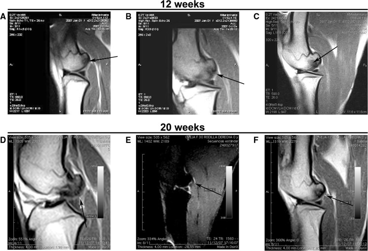
Fig. 2.
MRI images. The top images were taken at 12 weeks after arthroscopy: a untreated knee (sample F12left). b Defect treated with cell-free PLGA scaffold (sample F6left), where an incomplete osteointegration of the implant is shown by an arrow; and c osteochondral defect treated with cell-seeded PLGA scaffold (sample F6right), where the arrow points to an oedema-like signal in the subchondral bone. Bottom images were taken from samples recovered 20 weeks post arthroscopy: d untreated knee (sample F17left), where the arrow points at the irregular articular surface. e Lesion treated with cell-free PLGA scaffold (sample F30right), where the arrow points to the erosion on the articular surface; and f lesion treated with cell-seeded PLGA scaffold (sample F17right), where the arrow indicates the continuity of the surface

