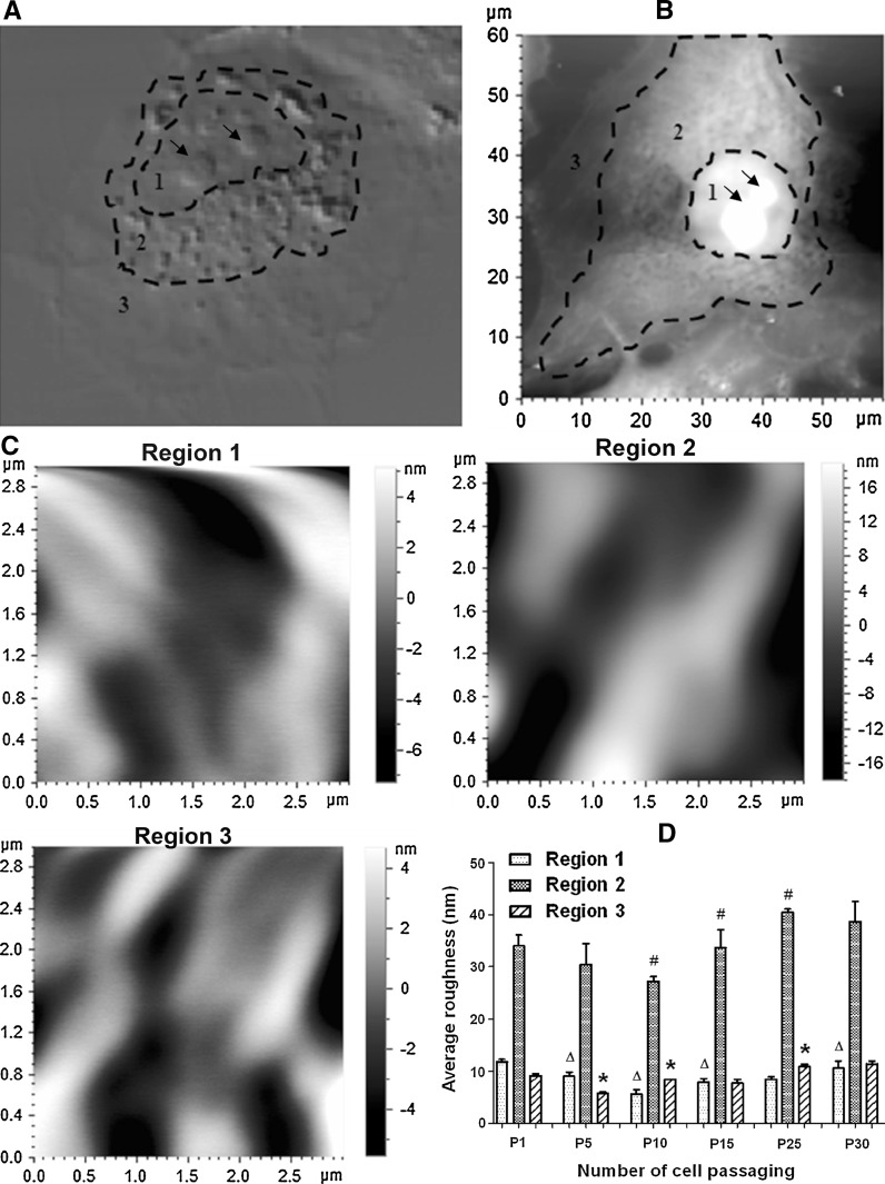
Fig. 4.
Effects of cell passaging on average cell-surface roughness of HUVECs. a A representative confocal DIC image shows the three regions with different degrees of roughness. b An AFM topographical image shows the three corresponding regions. c Representative AFM topographical images (3 μm × 3 μm) were enlarged from regions 1–3 of the cell in b, respectively. d The average roughness of each of the three regions on the cells at each passage (P1, P5, P10, P15, P25, or P30). During measurements of cell-surface roughness, the large protrusions in region 1 (as indicated by the arrows in a and b) were excluded since they were potentially caused by nucleoli. Values are expressed as mean ± SEM (n ≥ 6 from three independent experiments). triangle, ash, and asterisk correspond regions 1, 2, and 3, respectively, indicating the significant difference (P < 0.05) between a group and the previous group in average roughness of one of the three regions

