Fig. 2.

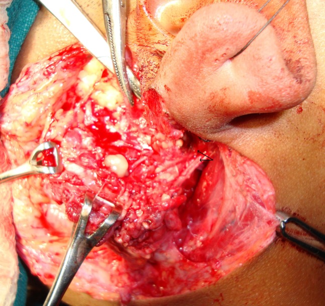
Pre-operative photograph depicting a branchial cyst in superficial lobe of parotid gland (white arrow) with aberrant facial (black arrow depicting two trunks of nerve coming out of stylomastoid foramen)

Pre-operative photograph depicting a branchial cyst in superficial lobe of parotid gland (white arrow) with aberrant facial (black arrow depicting two trunks of nerve coming out of stylomastoid foramen)