Figure 1.
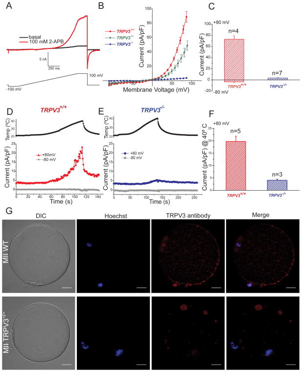
TRPV3 channels are expressed in MII mouse egg. A–F. Whole-cell voltage clamp recordings from an MII mouse egg. A. Current evoked from a voltage ramp from −100 to +100 mV in the absence (black trace) and presence (red trace) of 100 μM 2-APB. B. Current-voltage relations (I–Vs) in response to 100 μM 2-APB in WT (TrpV3+/+, CD-1 strain), heterozygous (TrpV3+/−) and eggs lacking TRPV3 (TrpV3-KO, TrpV3−/−). C. Averaged 2-APB evoked current recorded at +80 mV and −80 mV in WT and V3-KO eggs (73 ± 6 pA/pF for WT and 3.3 ± 0.5 pA/pF for V3-KO at +80 mV; −3.5 ± 0.8 pA/pF for WT and −0.2 ± 0.04 pA/pF for V3-KO eggs at −80 mV (± S.E.M). D–E. Temperature responses for WT and V3-KO eggs. Upper panels: Temperature (egg surface). Lower panels: Current responses at +80 mV (red trace) and −80 mV (gray trace) for WT and V3-KO eggs. F. Average TRPV3 current in response to 40°C recorded at +80 mV for WT and KO eggs. Current was 20 ± 2 pA/pF in WT eggs in contrast to only 4 ± 0.4 pA/pF in V3-KO eggs. The averaged inward current at −80 mV was −0.6 ± 0.2 pA/pF for WT and −0.4 ± 0.08 pA/pF for V3-KO eggs (data not shown). ± S.E.M; # of experiments are indicated over the bars. G. TRPV3 protein localization in mature mouse MII zona-free eggs; shown are differential interference contrast (DIC), DNA staining (Hoechst), and anti-TRPV3 staining. Upper panel: WT (129SvEvTac strain), Lower panel: TrpV3-KO egg. Most of the antibody-stained protein is at the membrane, except at the animal pole. Scale bar: 10 μm.

