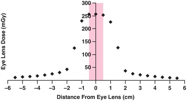
Fig. 4.

Graph shows eye lens dose as function of scanning location from protocol of American Association of Physicists in Medicine (80 kVp, 24 × 1.2-mm collimation, 270 mAs/rotation, 40 rotations) for brain perfusion CT examination using Sensation 64 scanner (Siemens Healthcare). Width of eye lens in z direction for this patient model (Irene, GSF [now Helmholtz Zentrum München]) is 1 cm. Shaded box indicates eye lens range in longitudinal direction. Therefore, when scanning location is 2 cm from center of eyes, eye lenses are completely out of x-ray beam (larger than half of beam width plus half of eye lens width).
