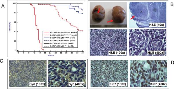
Fig. 2. Medulloblastomas in conditional BCCIP knockdown & p53 knockout mice.
The LoxPshBCCIP conditional BCCIP knockdown (BCCIP-CKD) mice were crossed with conditional p53 knockout (p53LoxP/LoxP) mice, and then with the GFAP-Cre mice that expressed the Cre recombinase during embryogenesis. This resulted in mice with 6 different genotypes as shown in the insert of panel A and Table 1. These mice were observed for tumor formation for over 48 months. Panel A shows the survival curve of the mice. Medulloblastomas were formed in 100% of the BCCIP-CKD;p53LoxP/LoxP mice with an average onset of 95 days. None of the other mice developed medulloblastoma. Some of the BCCIP-CON;p53LoxP/LoxP mice were sacrificed due to growth of benign lipomas at older ages (see text for details). Panel B shows the gross morphology of medulloblastoma formed in the BCCIP & p53 deficient BCCIP-CKD;p53LoxP/LoxP mice, and H&E staining of the representative tumors. The top left inserts show the medulloblastoma (indicated by arrows) from two mouse brains. The top-right insert shows that the tumor foci are within the cerebellar external granule layer with a typical histology of medulloblastoma. The bottom inserts are H&E staining of the tissues at the magnifications as indicated.
Panels C and D are IHC staining of the tumor tissue for synaptophysin (a medulloblastoma marker) and Ki67 (a proliferation marker).

