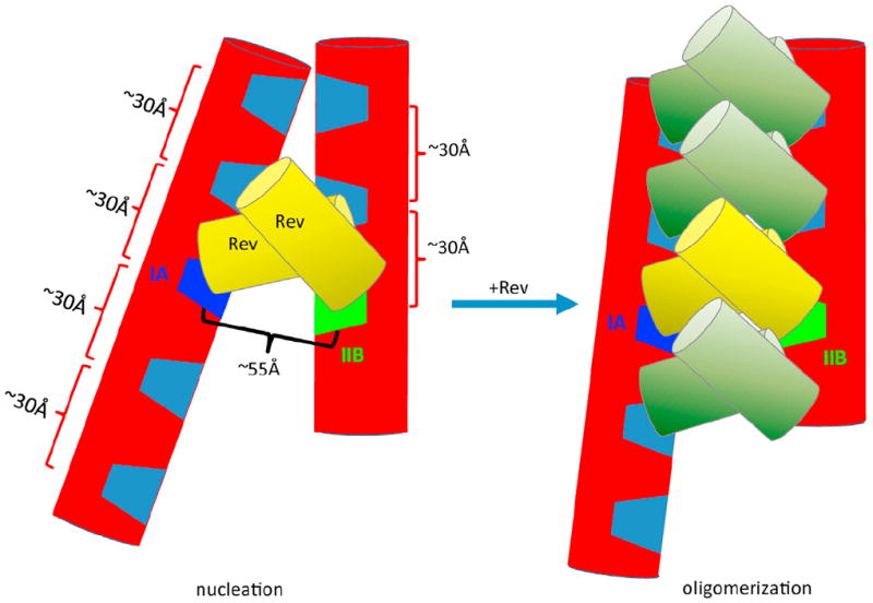
Figure 6. Models for Initial Rev Binding and Rev Oligomerization on the RRE RNA.

The RRE RNA is depicted in red cylinders with the IIB and IA Rev-binding sites highlighted in green and dark blue, respectively, and other major grooves shown in light blue. Initial binding of the first two Rev molecules (depicted in yellow cylinders) to the IIB and IA sites results in a nucleation site (left) for subsequent Rev oligomerization on the RRE RNA (right). This oligomerization is partially driven by hydrophobic interactions between the two Rev dimers and is constrained by the major groove spacing and the topological arrangement of the two major segments of the RRE. This oligomerization model illustrates the maximum number of Rev molecules that could potentially bind to this 233 nt RRE molecule based on spatial constraints. See also Figure S5 for a more realistic structural model.
