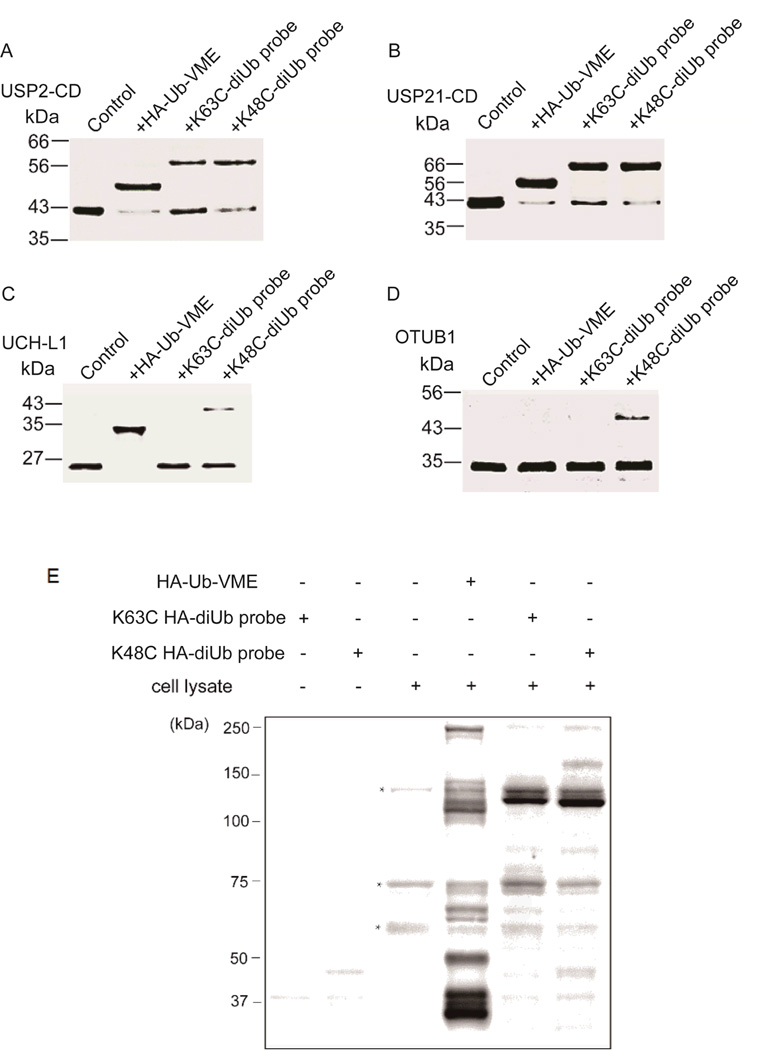
Fig. 2.
The reactivity of diUb probes towards different DUBs and DUB profiling using the diUb probes in cell lysates. (A-D) USP2-catalytic domain (USP2-CD), USP21-catalytic domain (USP21-CD), UCH-L1 and OTUB1 were incubated with different probes for 2 hr, separated by SDS-PAGE, and stained with Coomassie brilliant blue for detection. (E) DUB profiling using the diUb probes in cells in comparison to the monoubiquitin probe HA-Ub-VME. HEK293T cell lysates were labeled with HA-Ub-VME, K63C-linked HA-diUb probe and K48C-linked HA-diUb probe respectively. Samples were separated by SDS-PAGE and immunoblotted using anti-HA antibody. Asterisks indicate non-specific binding by the anti-HA antibody in cell lysate.

