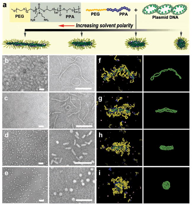
Figure 1.
Assembly of micellar nanoparticles with different shapes by condensing DNA with PEG10K-b-PPA4K block copolymer in solvents with different polarities. (a) Structure of PEG10K-b-PPA4K and schematic illustration of its assembly with DNA. (b–e) Morphologies of PEG-b-PPA/DNA micelles prepared in deionized water (b), 3:7 (v/v) (c), 5:5 (v/v) (d), and 7:3 (v/v) (e) DMF–water mixtures at an N/P ratio (ratio of primary amino groups in PPA block of the copolymer to phosphate groups in DNA) of 8. All scale bars represent 200 nm. (f–i) Representative images of PEG-b-PPA/DNA micelles obtained in molecular dynamics simulations corresponding to the conditions shown in panels (b–e). DNA is represented in green and the PEG and PPA blocks in yellow and blue, respectively. Monovalent counterions are depicted in pink. For clarity, the panels in the right-most column show the conformations of the plasmid DNA within the corresponding micelles depicted in panels (f–i).

