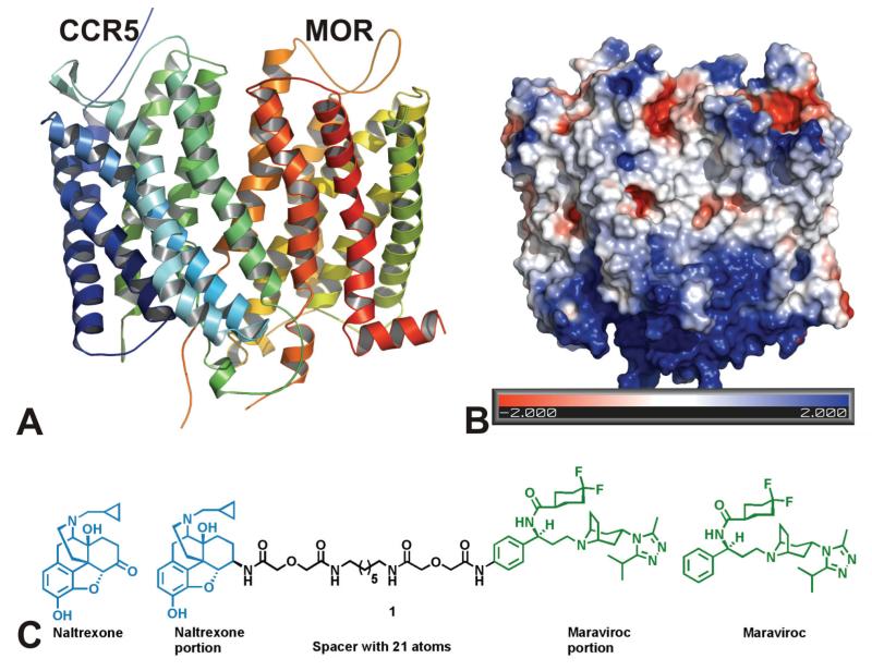
Figure 1. (A) Construction of a MOR-CCR5 dimer model.
Left: The ribbons colored in blue and green represent CCR5, and the ribbons colored in red and yellow represent MOR. Each ribbon was given an arbitrary color in order to distinguish each individual helix from the other. Right: Representation of the Poisson-Boltzmann electrostatic potentials at the surface of the heterodimer using the APBS plugin by PYMOL. Red represents acidic residues (−2 kBT/e); blue represents basic residues (+2 kBT/e); white represents uncharged residues. The figure indicates that the majority of the interactions between the two receptors are likely to be hydrophobic. (B) Construction of a chemical probe that interacts with both the MOR and CCR5 receptors simultaneously.

