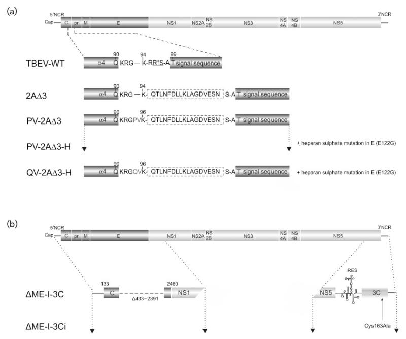
Fig. 1.
Structural organization of flavivirus mutants. (a) Drawing of the TBEV full-length genome and expanded views of the protein C of TBEV WT and each TBEV mutant (not drawn to scale). The NS2B/3 cleavage site is underlined and the cleavage position is marked by an asterisk. The inserted FMDV 2AΔ3 protein sequence is surrounded by a dashed line. The amino acids PV and QV preceding residue K96 were inserted to optimize the FMDV 3Cpro cleavage sequence. The numbers at the top refer to the amino acid positions within protein C. (b) Diagram of the TBEV genome and an expanded view of the structural protein region and the 3′NCR of each constructed TBEV replicon. The 3Cpro sequence was amplified by PCR from the FMDV strain O/SAR/19/00 and placed behind an EMCV IRES as described in methods. Engineered mutations are shown with the corresponding designation.

