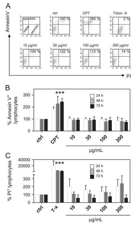
Fig. 2. Influence of Viola tricolor on lymphocyte apoptosis and necrosis.
Lymphocytes (2 × 10⁵ cells) were activated with PHA-L (10 μg/ml) alone or in the presence of camptothecin (CPT; 30 μg/mL), Triton-X 100 (0.5%) or different concentrations of the Viola extract (10–300 μg/mL) for 24 (white bars), 48 (gray bars) and 72 h (black bars). The amount of apoptotic (annexin V+/PI− and annexin V+/PI−) cells and necrotic cells (annexin V−/PI+) were assessed by FACS analysis using annexin V and propidium iodide staining. Representative dot plots and proliferation values (%) of total lymphocytes are demonstrated in (A). Results from total lymphocyte analysis of apoptotic (B) and necrotic (C) cells are summarized and presented as mean ± SD of three to four independent experiments. The values are normalized (=100%) to untreated stimulated cells (ctrl). The asterisks represent significant differences of Viola extract-treated cells in comparison to control cells (***P<0.001).

