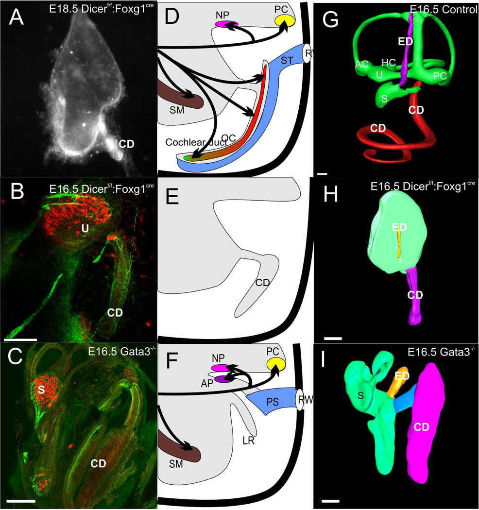
Figure 3.
This figure displays the loss of neurosensory development in the cochlea duct (CD) of two mutant mouse lines, a conditional deletion of Dicer 1 with Foxg1-cre (A,B) and deletion of Gata3 (C). Note that in either case hair cells and innervation exists to vestibular epithelia interpreted as utricle (U) and saccule (S), identifiable by the endolymphatic duct (ED) emanating from it. The cochlear duct is devoid of both hair cells and innervation other than autonomic fibers. The middle panel shows a diagram of the normal innervation and organization of the organ of Corti and the cochlear duct with the scala tympani (ST) attached (D), the loss of any innervation and hair cell formation in the two mutant lines but retention of the cochlear duct (E) and the loss of the basilar papilla and lagenar macula in a caecilian (F). The right column shows 3D reconstruction of confocal images of an E16.5 wildtype ear with Amira software (G), the remaining ‘ear’ of a conditional deletion of Dicer with the cochlear duct extending from the undivided upper part (H) and the partially developed ear of a Gata3 null mouse with a cochlear duct connected via a ductus reunions to the vestibular part of the ear. For abbreviations see Fig. 2. Images taken from (Duncan et al. 2011; Kersigo et al. 2011).

