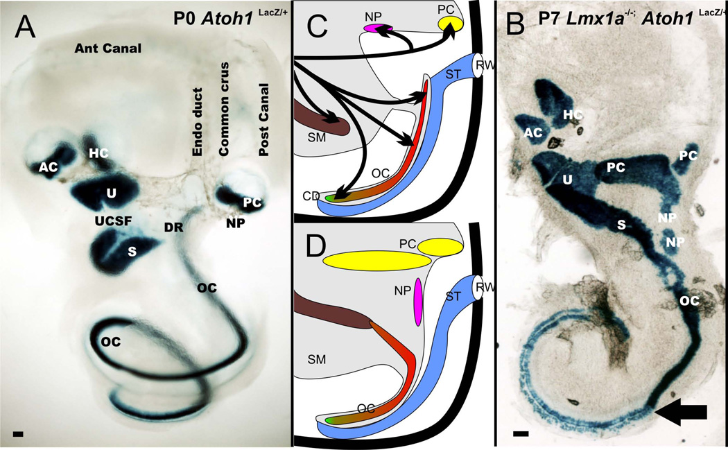
Figure 4.
The distribution of the six mammalian sensory epithelia and the neglected papilla (NP) is shown for a wildtype ear (A) and Lmx1a mutant ear (B). Note that there is continuity of utricle (U), saccule (S), and organ of Corti (OC) in the mutants. Also, the cochlea has a basal part that is converted into a more vestibular like system and an apex that is more cochlear like indicating different effects of Lmx1a along the length of the cochlea. There is a great enlargement of the posterior canal crista as well as the two patches of neglected papilla (NP) extending into the wide cochlear duct. The middle column shows the transformation of the ear in the Lmx1a null mutant. The saccular macula fuses with the organ of Corti, and there is no approximation of the scala tympani with the base of the organ of Corti but only with the apex (D). There is a dramatic increase in size of the posterior canal crista and neglected papilla. AC, anterior canal crista; HC, horizontal canal crista; DR, ductus reuniens; RW, round window; SM, scala media; UCSF; utriculo-saccular foramen. Modified after (Nichols et al. 2008)

