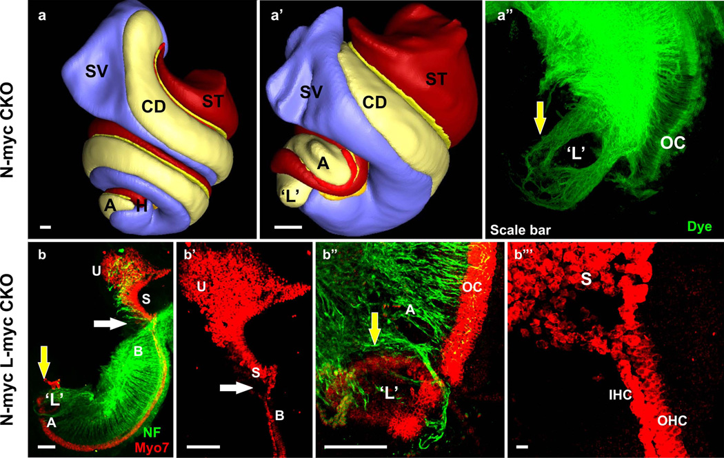
Figure 5.
Three dimensional reconstruction of the inner ear shows the different regions of the cochlea in wildtype (a). Deletion of N-Myc results in severe truncation of the cochlear duct and aberration in the appropriate segregation of the different regions of the cochlea (b). In particular, the apex of the N-Myc CKO cochlea forms a circular sac-like structure resembling a ‘lagena’ (yellow arrow in b’). Dye tracing from the cochlear nucleus shows afferent innervation to the abnormally formed circular sac or ‘transformed lagena’. Double knockout (dCKO) of N-Myc and L-Myc adds to the severity in the N-Myc CKO phenotype (c-c”‘). Myo7a immunohistochemistry shows abnormal fusion of the hair cells from the basal tip with the saccular hair cells (white arrows in c-c”). The dCKO also shows formation of the segregated circular sac from the apical tip of the cochlea (yellow arrows in c, c”‘). The shortened cochlea forms multiple rows of hair cells in the apex (c”). Neurofilament immunohistochemistry shows the overshooting of the fibers to this circular sac (c”‘). A, apex; B, base; CD, cochlear duct; L, lagena; OC, organ of Corti; S, saccule; SV, scala vestibuli; ST, scala tympani; U, utricle. Bar indicates 100 µm. Modified after (Kopecky et al. 2011; Pan et al. 2011; Kopecky et al. 2012).

