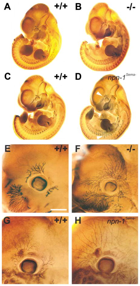
Figure 2. Peripheral Projections of Cranial and Spinal Nerves Are Severely Disorganized in Both npn-1 Null and npn-1Sema− Mice.
(A–D) Whole-mount antineurofilament staining of E11.5 npn-1 null (B), wild-type littermate (A), homozygous npn-1Sema− (D), and wild-type littermate (C) embryos. The ophthalmic branch of the trigeminal nerve (upper arrowhead in [D]) and spinal nerves (lower arrowhead in [D]) are disorganized in both npn-1 null (B) and npn-1Sema− mice (D). (E–H) Whole-mount antineurofilament staining of the ophthalmic nerve in E12.5 homozygous npn-1 null (F), wild-type littermate (E), homozygous npn-1Sema− (H), and wild-type littermate (G) embryos. Note the exuberant extension and more regular distribution of ophthalmic nerve branches in npn-1Sema− as compared to npn-1 null ophthalmic projections. Scale bar: (A–D), 0.5 mm; (E–H), 0.2 mm.

