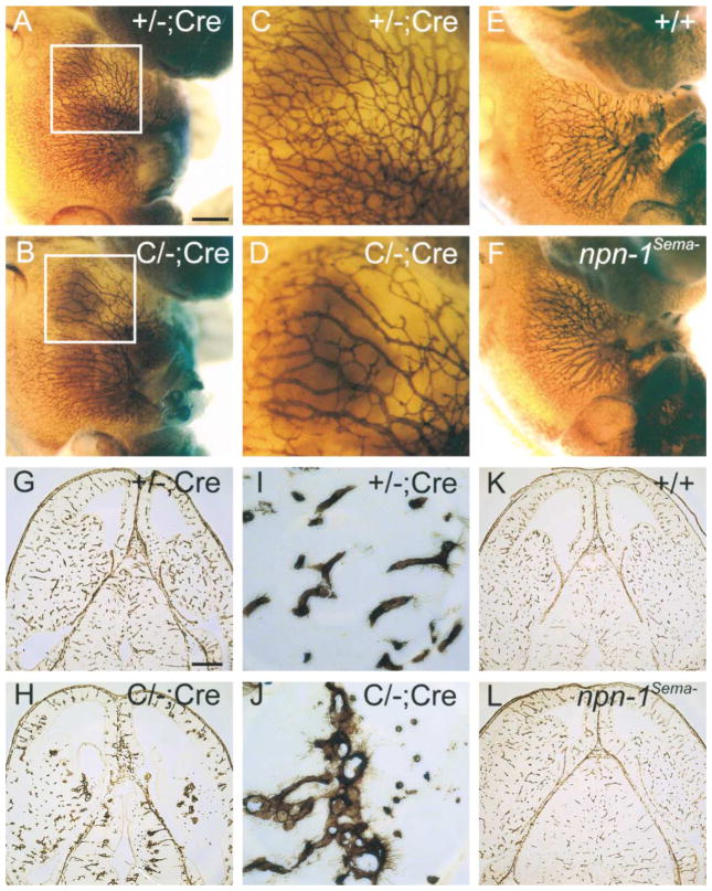
Figure 6. Severe Vasculature Disruption in Endothelial-Specific npn-1 Null Mice, but Not in npn-1Sema− Mice.
(A–F) Whole-mount anti-PECAM staining of a endothelial cell-specific npn-1 null mutant (C/−; Cre) (B), a heterozygous littermate control (+/−; Cre) (A), homozygous npn-1Sema− (F), and a wild-type littermate control embryo (E), all at E12.5. Boxed regions from (A) and (B) are displayed at higher magnification in (C) and (D), respectively.
(G–L) Isolectin staining of E13.5 horizontal brain sections from a endothelial cell-specific npn-1 null mutant embryo (C/−; Cre) (H), a heterozygous littermate control embryo (+/−;Cre) (G), a homozygous npn-1Sema− embryo (L), and a wild-type littermate control embryo (K). Select regions from (G) and (H) are displayed at higher magnification in (I) and (J), respectively. Note the decrease in endothelial branching in C/−; Cre mice compared to controls. Scale bars: (A, B, E, and F), 0.5 mm; (C and D), 0.2 mm; (G, H, K, and L), 350 μm; (I and J), 50 μm.

