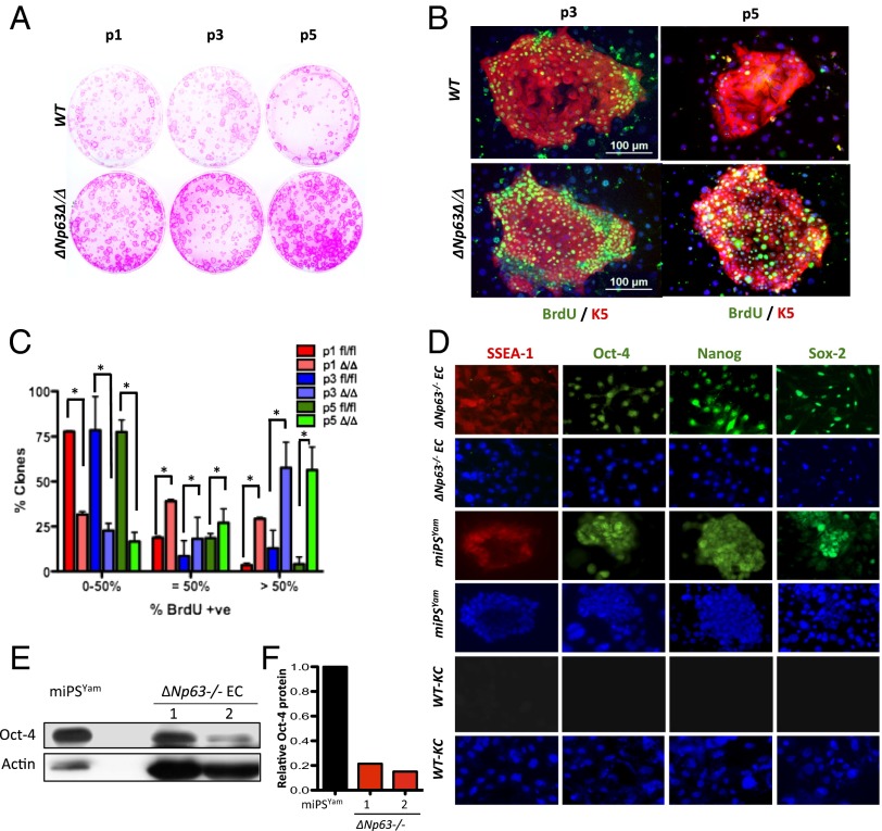
Fig. 1.
ΔNp63-deficient epidermal cells are proliferative. (A) Epidermal colonies of the indicated genotypes stained with rhodamine B. Passages 1 (P1), 3 (P3), and 5 (P5) are shown. (B) Immunostaining for BrdU (green) and K5 (red) in P3 and P5 epidermal cells. (C) Quantification of BrdU incorporation in P1, P3, and P5 colonies after 8 d in culture. Asterisks indicate statistical significance (P < 0.001). (D) IF performed on the indicated cells using the indicated antibodies. (E) Western blot performed on lysates from miPSYam and ΔNp63−/− cells derived from two independent embryos (1 and 2) using the indicated antibodies. Actin was used as loading control. (F) Quantification of the Western blot in E.

